Chest X Rays Showing Pneumonia

When a clinician reviews chest x rays showing pneumonia, the image often reveals telltale shadows that signal infection and guide the next steps in care. Pneumonia can appear in many forms on a chest radiograph, and understanding these patterns helps providers confirm the diagnosis, track progression, and choose the right treatment. From the first view to follow up scans, the way the lungs look on each image tells a story about inflammation, fluid, and the body’s response.
How Pneumonia Appears on a Chest X Ray
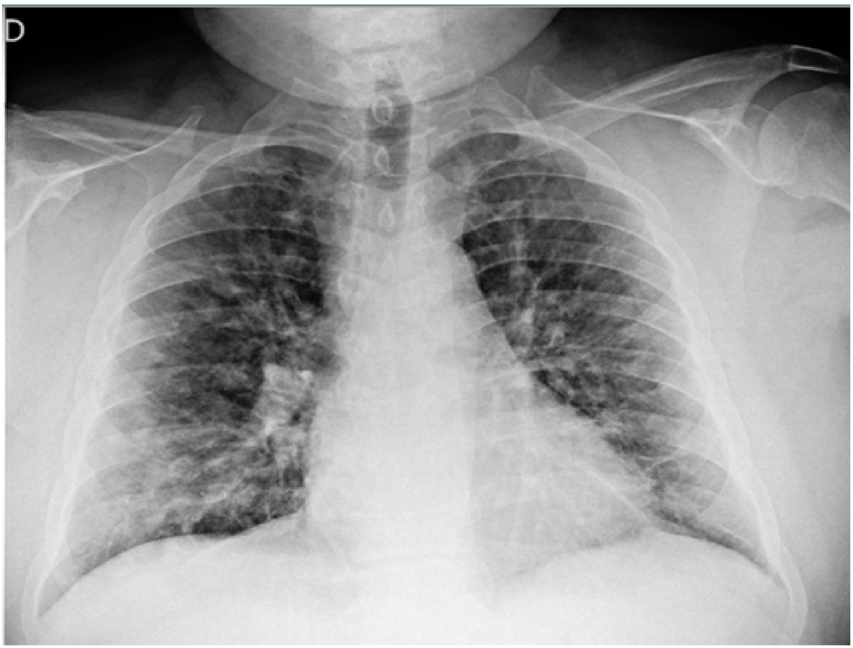
On a typical chest x ray, pneumonia often shows as areas of increased opacity where infection has settled in the lung tissue. These cloudy or white regions may look lobar, covering a whole section of the lung, or more patchy and scattered when the illness affects multiple areas. The pattern you see depends on the type of germ, how deeply it has spread, and whether the infection is just beginning or has been present for some time. A careful reader of chest x rays showing pneumonia will also notice differences in density, air bronchograms, and how clearly the edges of the heart and diaphragm remain visible.
An air bronchogram, where air-filled bronchi stand out against infected, fluid-filled lung, is one classic clue that helps distinguish pneumonia from other causes of opacity. Sometimes the image shows subtle changes at the lung bases, while in other cases one entire lower lobe appears heavily involved. Because many conditions can mimic these findings, clinicians compare the current study with older images, review the patient’s symptoms, and consider risk factors to be confident about the diagnosis. This careful approach reduces the chance of mistaking atelectasis, fluid in the space around the lung, or even a tumor for active infection.

Lobar Versus Bronchopneumonia Patterns
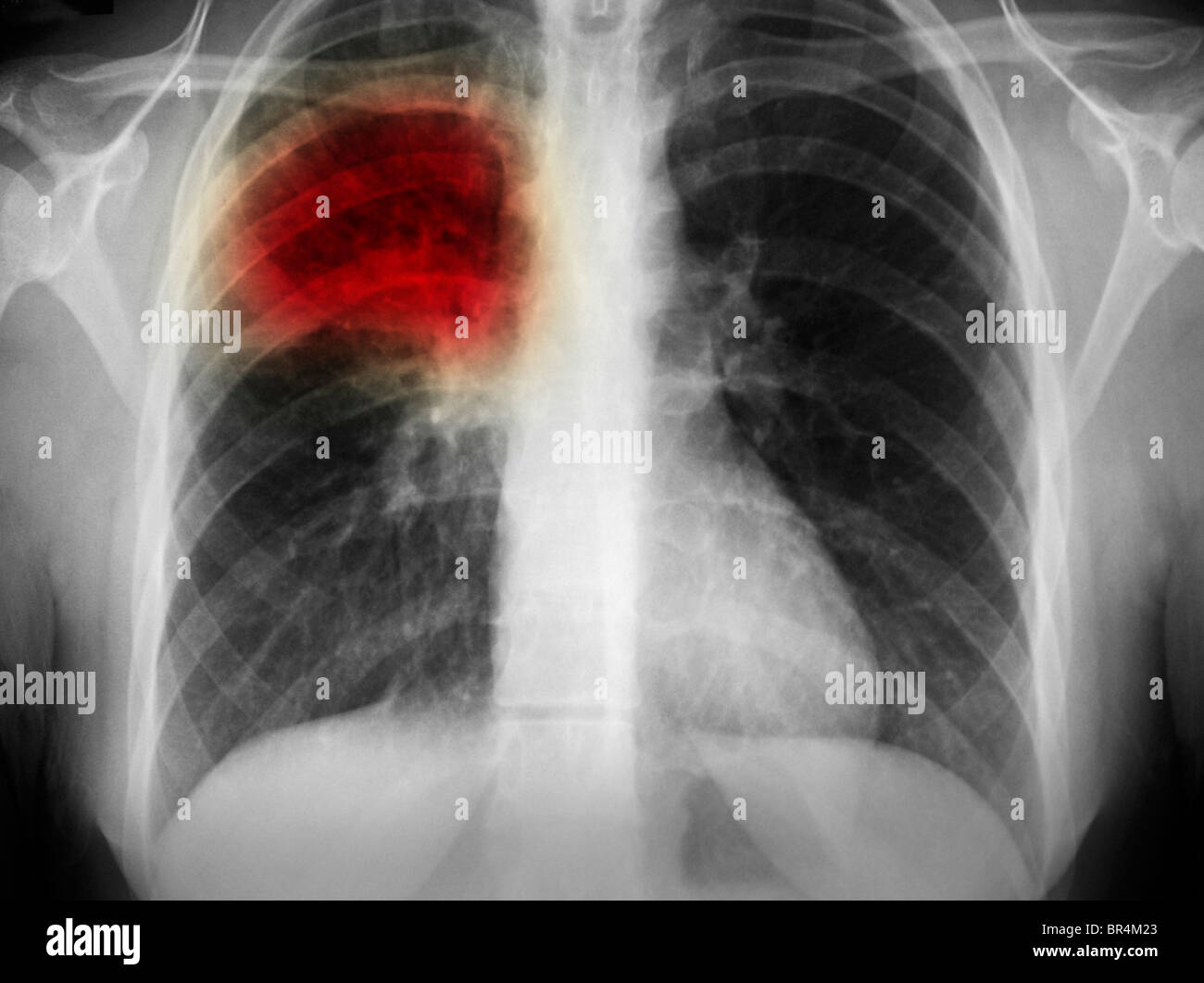
Lobar pneumonia tends to fill an entire lung segment or lobe with dense consolidation, so the affected area on the chest x ray looks like a solid, well-defined zone of opacity with a visible edge. In contrast, bronchopneumonia often appears as scattered patches centered around the airways, creating a more patchwork pattern across both lungs. Recognizing these patterns on chest x rays showing pneumonia helps narrow down the likely causes and guides decisions about hospitalization, antibiotics, and supportive care.
When the infection is lobar, the involved lobe may also appear slightly larger than usual, and the surrounding lung often shows subtle changes as the body responds. With bronchopneumonia, the patchy infiltrates can move or change quickly, especially if the infection is severe or the patient is immunocompromised. Radiologists and clinicians pay close attention to the distribution, because these differences influence how quickly the patient is likely to improve and whether additional imaging or tests are needed.
Complications and Atypical Findings
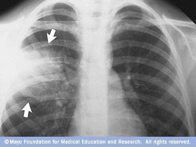
Not every chest x ray showing pneumonia looks textbook, and sometimes the images reveal complications that demand extra attention. For example, an abscess may appear as a cavity with an air-fluid level, while empyema shows up as a thickened, irregular pleural line and fluid that collects around the lung. These findings change how clinicians manage the illness, often requiring longer antibiotic courses, drainage procedures, or closer monitoring to ensure the infection is resolving safely.
- Abscess formation with air-fluid levels inside a cavity.
- Empyema, or pus in the space between the lung and chest wall.
- Pleural thickening or effusion seen as hazy, dense areas near the edges of the lung.
In older adults, people with weakened immune systems, or those with other lung diseases, the signs on chest x rays showing pneumonia can be subtler. Instead of classic dense consolidation, the image may show only subtle increases in opacity, atelectasis, or a slightly hazy lung base. This is why clinicians combine imaging with a careful physical exam, lab tests, and sometimes additional scans to be sure they are not missing early or atypical disease.
Comparing Current and Prior Images
One of the most powerful tools in interpreting chest x rays showing pneumonia is comparing the current view with previous studies. When an area that was once clear now appears cloudy, or when a hazy zone grows larger, it strongly supports a new or worsening infection. By contrast, stable findings over weeks or months suggest that the opacity may be due to scarring, old infection, or another non-infectious cause.
Serial imaging also helps clinicians see how well treatment is working. If the white areas on the x ray begin to clear and the lung edges sharpen, that is a reassuring sign that the antibiotics or other therapies are doing their job. On the other hand, persistent or expanding changes may prompt a reevaluation of the diagnosis, a switch in medication, or further testing to rule out unusual pathogens or complications.
When Additional Tests Are Needed
Although chest x rays showing pneumonia are often enough to start treatment, there are times when extra information is necessary. A computed tomography (CT) scan can provide more detail, especially when the plain film is unclear, complications are suspected, or the patient does not improve as expected. CT images can highlight small abscesses, subtle infiltrates, or patterns that suggest a particular cause, such as a viral infection or an unusual organism.
Laboratory results, cultures, and clinical context also shape how clinicians use imaging. Blood tests, sputum samples, and sometimes bronchoscopy with samples taken directly from the lung help confirm the germ and refine treatment. In this way, chest x rays showing pneumonia act as a bridge between the patient’s symptoms and the targeted therapy that will speed recovery.
Follow-Up and Long-Term Outlook
After treatment, follow-up chest x rays are commonly used to confirm that the pneumonia has fully resolved, especially in people who were hospitalized or had severe disease. In many cases, a repeat image weeks later shows clear lungs, with no lingering signs of the earlier infection. However, some patients, particularly those with slow recovery or underlying lung problems, may still have faint haziness for a longer period.
For most people, a single episode of pneumonia that is diagnosed promptly and treated appropriately leads to a full recovery, and the final images look essentially normal. For others, especially older adults or those with chronic conditions, repeated episodes or slow clearing of chest x rays showing pneumonia may point to broader issues such as weakened airways, immune problems, or structural lung disease. Working closely with clinicians, using smoking cessation when relevant, and staying up to date with vaccines can lower the risk of future problems and help each new episode be caught and managed early.
Chest x rays showing pneumonia remain a cornerstone of diagnosis and follow-up, providing a clear visual map of where infection is located and how it is responding to care. By combining these images with clinical judgment, lab results, and patient history, clinicians can guide treatment, watch for complications, and support long-term lung health.
🚨Chest X-Ray Findings in Pneumonia with Pleural Effusion 🩻 #shorts #radiology #chest #doctor
In this short, we analyze a chest X-ray showing pneumonia with a pleural effusion—a key radiographic sign indicating fluid ...
