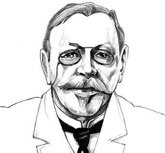
Hans Christian Joachim Gram

Hans Christian Joachim Gram shaped the foundation of modern microbiology with a simple yet revolutionary staining technique that still defines how we identify bacteria today. The Gram stain remains a cornerstone in clinical laboratories around the world, guiding treatment decisions and helping clinicians distinguish between two major categories of bacteria. This method emerged from the careful work of Gram, a Danish scientist whose curiosity and meticulous approach turned a routine staining experiment into a lasting diagnostic tool. Understanding his background, the historical context, and the technical details of the procedure reveals why the Gram stain continues to be so influential more than a century later.
Early Life and Scientific Training
Hans Christian Joachim Gram was born in Copenhagen, Denmark, in 1853, growing up in an environment that encouraged intellectual exploration and academic rigor. He studied medicine at the University of Copenhagen, where he built a solid foundation in anatomy, physiology, and the emerging sciences of microbiology and pathology. During his formative years, the field of bacteriology was rapidly expanding, driven by discoveries from pioneers such as Pasteur and Koch, who demonstrated the role of microorganisms in disease. Gram trained under the influential scientist Carl Friedländer, and it was during his work in pathological anatomy that he encountered the practical challenges of identifying bacteria in tissue samples. These early experiences shaped his attention to detail and laid the groundwork for the staining method that would eventually bear his name.
After completing his medical doctorate, Gram worked at several hospitals and research institutions, refining his skills in microscopy and experimental techniques. The scientific community of the late nineteenth century was intensely focused on classifying microorganisms and understanding their structural differences, yet reliable methods were still limited. Gram recognized that existing staining procedures did not reliably differentiate between types of bacteria, which often led to ambiguous or inconsistent results in clinical settings. By combining insights from chemistry, dye properties, and microscopic observation, he set out to develop a more systematic approach. This pursuit of clarity and precision ultimately led to the publication of his now-famous staining procedure in 1884, establishing a standard that would endure in both teaching laboratories and diagnostic departments.

Discovery of the Gram Stain Technique
The discovery of the Gram stain technique emerged from careful experimentation rather than a single dramatic breakthrough. Gram was investigating bacterial samples in the context of lung infections when he observed that certain bacteria retained a violet dye after a decolorization step, while others lost the color and appeared pink after counterstaining. This difference in staining behavior reflected fundamental variations in bacterial cell wall structure, although the precise biochemical explanations would come later. Gram documented these observations methodically, testing variations in dye concentration, timing, and the sequence of staining steps. His initial report in 1884 outlined a simple yet powerful procedure that could reliably separate bacteria into two broad groups, which later became known as Gram-positive and Gram-negative organisms.
Key elements of the original Gram method included crystal violet as the primary stain, iodine as a mordant to form a stable complex, alcohol or acetone for decolorization, and a counterstain such as safranin to visualize the decolorized cells. The interaction between these reagents and the bacterial cell wall depends on the thickness of the peptidoglycan layer and the presence of an outer membrane, concepts that were not fully understood at the time but have since been clarified by advances in molecular biology. Gram’s approach emphasized reproducibility, showing that careful control of each step produced consistent results across different laboratories. This robustness helped the technique spread quickly and become integrated into standard microbiological practice, where it remains a fundamental skill for students and professionals alike.
How the Gram Stain Works: Basic Principles
The Gram stain operates through a sequence of steps that highlight structural differences in bacterial cell walls. After fixing bacteria to a slide and applying the primary stain crystal violet, all cells appear purple under the microscope. The addition of iodine traps the dye within the cell by forming large complexes, enhancing retention during the subsequent washing step. When a decolorizing agent such as alcohol is applied, two distinct outcomes emerge. Gram-positive bacteria, with their thick layer of peptidoglycan, retain the crystal violet-iodine complex and remain purple, while Gram-negative bacteria, which have a thinner peptidoglycan layer and an outer membrane, lose the dye and become colorless. The final application of a counterstain, typically safranin, then colors these decolorized cells pink, providing clear visual contrast between the two groups.

Although the procedure appears straightforward, subtle variations in reagents, timing, and technique can influence the results, making quality control essential in clinical and research settings. Heat fixation, the concentration of dyes, and the duration of decolorization all play critical roles in ensuring accurate classification. Understanding these factors helps explain why the Gram stain continues to be taught with precision and why laboratories follow strict protocols. For students and practitioners, mastering the Gram stain offers not only a practical skill but also an insight into the structural diversity of bacteria and the historical development of microbiological methods.
Impact on Medicine and Clinical Practice
In medicine, the Gram stain provides rapid, preliminary information about the type of bacteria potentially responsible for an infection, which can be crucial when immediate treatment decisions are required. By distinguishing between Gram-positive and Gram-negative pathogens, clinicians can narrow down the range of appropriate antibiotics before more specific identification tests are completed. For example, Gram-positive infections often respond well to certain beta-lactam antibiotics, while Gram-negative organisms may require drugs that can penetrate their outer membrane. This initial classification helps guide empirical therapy in settings such as emergency departments, intensive care units, and outpatient clinics, where timely intervention is essential.
Beyond antibiotic selection, the Gram stain supports the identification of specimens from blood, sputum, urine, and other body fluids, contributing to the diagnosis of pneumonia, sepsis, meningitis, and many other conditions. Laboratories rely on the expertise of skilled technologists who perform the stain with consistent technique and interpret the results in combination with other findings. While molecular methods and mass spectrometry are increasingly used for precise identification, the Gram stain remains a fast, low-cost, and broadly accessible tool. Its continued relevance demonstrates how a nineteenth-century innovation can evolve alongside modern technology while maintaining a central role in patient care.
Legacy and Ongoing Relevance
The legacy of Hans Christian Joachim Gram extends far beyond the staining protocol that bears his name, influencing how generations of microbiologists think about bacterial classification and cell structure. The simplicity and elegance of the Gram stain have made it a symbol of scientific ingenuity, demonstrating how a carefully designed experiment can yield insights that last for decades. Educational curricula worldwide continue to include the Gram stain as a foundational technique, ensuring that new generations of health professionals appreciate both its historical significance and its practical utility. Research into bacterial pathogenesis, antibiotic development, and diagnostic innovation still references the Gram classification system, highlighting its enduring conceptual importance.
Today, the principles underlying the Gram stain inform more advanced technologies, from automated imaging systems to molecular assays that target cell wall components. Variations of the original method have been adapted for specific applications, including fluorescent stains and simplified protocols for resource-limited settings. As antimicrobial resistance becomes an increasingly critical global health issue, the ability to quickly categorize bacteria remains valuable for guiding appropriate therapy and public health responses. Hans Christian Joachim Gram’s contribution thus lives on not only in textbooks and laboratories but also in the everyday practice of medicine, where his careful observations continue to shape how we understand and treat bacterial infections.
Hans Christian Joachim Gram bacteoriólogo
Hans Christian Joachim Gram fue un bacteriólogo danés que desarrolló la tinción de Gram, un método de amplio uso en ...
