X Rays Of Fractured Ankle

When someone experiences a sudden twist, fall, or blow to the lower leg, one of the first diagnostic tools a clinician turns to is imaging of the area, and x rays of fractured ankle are often the initial step in assessing the severity of the injury.
Understanding the Ankle Anatomy and Injury Mechanism
The ankle joint is a complex hinge formed by the tibia, fibula, and talus, held together by strong ligaments that control movement and provide stability. A fracture can occur in any of these bones when the forces applied to the joint exceed the strength of the surrounding structures, such as during a sports injury, a fall from height, or a motor vehicle accident.
Because the ankle bears the entire weight of the body with every step, even a hairline crack can significantly impair mobility and cause substantial pain. Recognizing the type and location of the break is essential for determining the correct treatment plan, and this is where x rays of fractured ankle imaging plays a critical role in guiding the next steps toward recovery.
How X Rays Capture the Injury
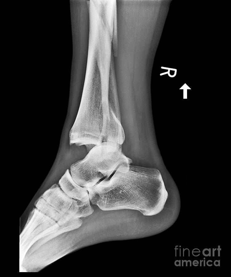
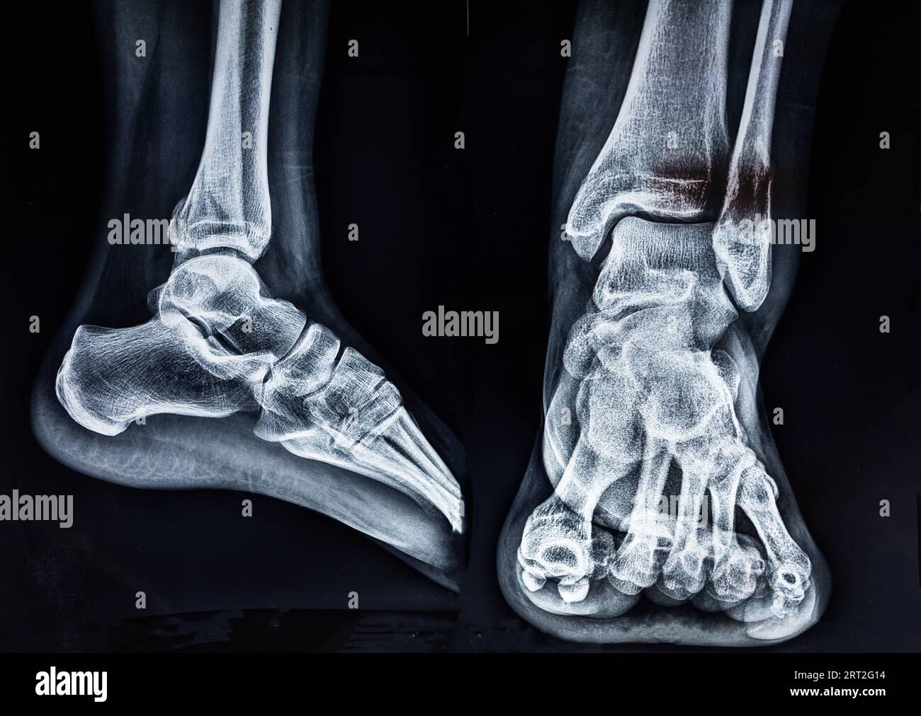
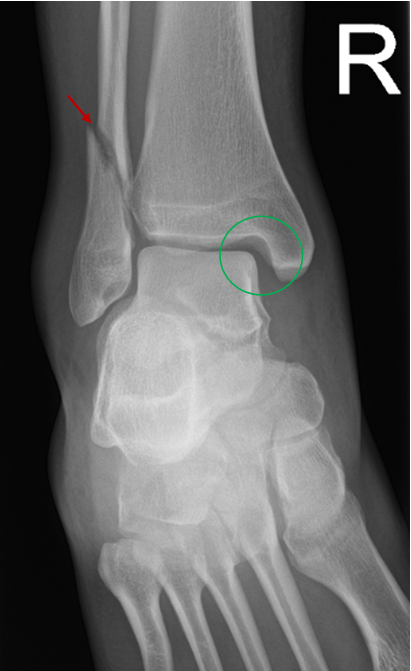
An x ray uses a small amount of focused radiation to create a two-dimensional image of dense structures like bone, allowing the radiologist and physician to see the exact alignment of the bones in the joint. In the case of x rays of fractured ankle, the images will reveal whether the crack is clean and non-displaced, or if the bone fragments have shifted out of their normal position.
During the procedure, the patient typically stands or sits while a specialized machine directs the beam through the ankle from different angles. The resulting picture, or radiograph, shows the bones in shades of white against a darker background, making it possible to identify fracture lines, joint spaces, and any potential dislocation with high clarity.
Interpreting the Radiograph Results
Reading x rays of fractured ankle requires a trained eye to differentiate between stable and unstable patterns. A stable fracture means the bone pieces remain aligned, while an unstable fracture indicates that the fragments may move out of place, often necessitating surgical intervention to restore proper function.
- Non-displaced fractures appear as a thin line across the bone with no visible gap or shifting of the segments.
- Displaced fractures show clear separation or angulation of the bone pieces, which can compromise the stability of the joint.
- Comminuted fractures reveal the bone broken into several pieces, which can complicate healing and require precise surgical repair.
Complementary Imaging and Advanced Diagnostics
While x rays of fractured ankle are excellent for visualizing bone, they do have limitations when it comes to soft tissue structures like ligaments, tendons, and cartilage. If the clinical exam suggests damage beyond the bone, a physician may order additional imaging to get a fuller picture of the injury.
In these situations, more advanced modalities such as MRI or CT scans may be used alongside the initial x ray results. These techniques provide high-resolution, three-dimensional views that help surgeons plan complex procedures and ensure that all damaged tissue is addressed during treatment.
Common Treatment Pathways Based on Imaging
The findings on x rays of fractured ankle directly influence the treatment strategy, ranging from conservative management to surgical repair. For minor breaks where the bones remain aligned, doctors often recommend immobilization using a cast or walking boot to allow the bone to heal naturally over several weeks.
When the fracture is displaced or involves the joint surface, realignment under anesthesia followed by surgical fixation with plates, screws, or rods may be necessary. The goal in every case is to restore the normal anatomy of the joint so that the patient can regain strength, stability, and a full range of motion without chronic pain or instability.
Recovery, Rehabilitation, and Long-Term Outlook
After the initial healing phase, rehabilitation becomes a cornerstone of recovery, especially when x rays of fractured ankle show that the bone alignment has been successfully restored. Physical therapy helps rebuild muscle strength, improve balance, and restore flexibility, reducing the risk of future injuries.
Most patients who follow their treatment plan diligently can expect a return to normal daily activities and, in many cases, a full return to sports and high-impact exercise. Consistent follow-up imaging may be used to monitor the healing process and ensure that the fracture has united properly without complications.

In conclusion, x rays of fractured ankle remain an indispensable tool in the rapid and accurate diagnosis of ankle injuries, providing essential information that guides every stage of care. By combining clear imaging with expert medical judgment and dedicated rehabilitation, individuals can achieve the best possible outcomes and get back to moving comfortably in their daily lives.
Ankle Fractures: Pathologic changes seen on radiographs
Ankle Fracture Educational Series - Part 2 of 3 Produced by faculty and trainees of the Harvard Global Orthopaedics Collaborative ...
